
Medically reviewed by
Assoc. Prof. Dr. Ayhan Işık Erdal, MD, FACS, FEBOPRAS
Plastic, Reconstructive & Aesthetic Surgeon
· Last reviewed
Bottoming out, lateral displacement, double bubble, symmastia. Whichever the pattern, the underlying problem is the same: the pocket has expanded beyond where it should be, and the implant has settled in the wrong position. The fix is internal pocket reconstruction.
An implant in the correct position sits where the surgeon designed it: within an inframammary fold of the right height, with the implant filling the breast mound, neither too medial nor too lateral. Malposition means the implant has moved away from this design — sometimes within weeks of surgery, sometimes years later.
Malposition is not the same as size mismatch. A correctly positioned but oversized implant will look wrong because of size, not position. A correctly sized implant in the wrong position will look wrong because of position. Distinguishing these matters for the surgical plan.
| Type | What it looks like | Cause |
|---|---|---|
| Bottoming out | Implant has dropped below the original fold. Nipple appears too high; lower pole stretched out. | Inframammary fold released too aggressively, weight of large implants over time, tissue laxity. |
| Lateral displacement | Implant has moved toward the armpit. Cleavage area looks empty when supine. | Lateral pocket dissected too far, or pocket has stretched laterally over time. |
| Symmastia ("uniboob") | Implants too close together at midline; cleavage gap is gone or reversed. | Medial pocket dissected too far, undermining sternal attachment. |
| Double bubble | Visible horizontal fold below the implant — the implant has descended below the original IMF, but the original IMF is still adherent. | Common in down-fold revisions, constricted breast revisions, or when implant is too large for the original IMF position. |
Visual examination plus measurements:
Comparing pre-primary photos (if available), early post-primary photos, and current photos shows the trajectory — was malposition immediate or progressive? This affects surgical planning.
The operation depends on type:
The expanded inferior pocket is reconstructed with internal capsulorrhaphy — permanent sutures placed inside the capsule to recreate the inframammary fold at the proper height. The pocket is reduced from below; the new fold is anchored to chest wall structures.
Lateral capsulorrhaphy reduces the pocket from the lateral side. Permanent sutures placed along the lateral capsule wall reduce pocket dimensions and prevent re-displacement.
Most challenging type. Medial capsulorrhaphy plus restoration of the sternal soft-tissue cover. May require ADM (acellular dermal matrix) reinforcement if tissue is very thin. Implant may need to be downsized.
The original adherent IMF must be released and the new fold raised to the proper height. Capsulorrhaphy at the new fold level.
The technical centrepiece of malposition repair. Permanent sutures placed inside the capsule reduce the pocket dimensions and reinforce key boundaries — particularly the inframammary fold. The repair is invisible from outside but holds the implant in the correct position long-term.
Three layers of sutures are typical: deep (capsule to chest wall structures), middle (capsule wall to capsule wall), superficial (subcutaneous reinforcement). Permanent monofilament sutures (typically Prolene) are used.
For complex or recurrent cases, a sheet of ADM or surgical mesh may be added internally to provide additional structural support. This adds cost but improves long-term outcomes in select cases.
Internal capsulorrhaphy recovery is longer than other revision types — the internal sutures need time to integrate with healing capsule. Resistance training too early can stress the repair.
Permanent sutures provide long-term reinforcement. The internal repair becomes part of the new capsule structure as healing occurs over 3-6 months. Stability beyond one year is excellent in well-performed cases — most failures occur in the first 3 months if they're going to occur.
Multiple factors: original pocket made too low, overly aggressive inframammary fold release, gravity acting on heavy implants over time, weakened tissue support, and mass effect of large implants stretching the lower pole.
Not always. For routine malposition repair, capsulorrhaphy with permanent sutures is usually sufficient. Acellular dermal matrix (ADM) or surgical mesh is reserved for complex cases — recurrent malposition, very thin tissue, or significant pocket distortion. ADM adds cost and complexity, so it's used only when indicated.
Yes. Asymmetric malposition (e.g., bottoming out on one side, lateral displacement on the other) is not uncommon and requires asymmetric repair. Each side is addressed according to its specific findings.
Free WhatsApp consultation in English with Dr. Erdal's clinic. Send your photos for an initial assessment.
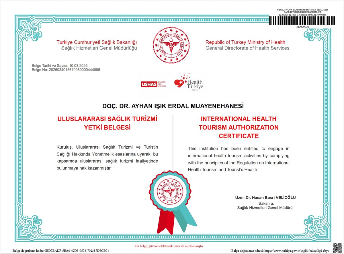
This clinic is officially authorised by the Republic of Türkiye Ministry of Health (Sağlık Bakanlığı, General Directorate of Health Services) to provide international health tourism services. The Ministry audits the clinic's surgeon credentials, facility standards, infection-control protocols, and complication-tracking systems before issuing this certification.
Click certificate to view at full resolution. Document carries digital signature and QR-code verification on the original.